Foot Muscles Mri Anatomy - - Tendinous, ligamentous, and muscle abnormalities.. Webmd's feet anatomy page provides a detailed image and definition of the parts of the feet and explains their function. The feet are flexible structures of bones, joints, muscles, and soft tissues that let us stand upright and perform activities like walking, running, and jumping. The muscles acting on the foot can be divided into two distinct groups; Neuropathies around the elbow joint. The muscles that control the movements of the foot originate in the lower leg and are attached the bones in the foot with tendons.
Almost every movement in the body is the outcome of muscle contraction. Radiologists perform ankle imaging to assess injuries of the foot and ankle anatomy. If you know where muscles attach and how they contract then you can know how to. Variants, accessory muscles and ossicles. Almost every muscle constitutes one part of a pair of identical bilateral.
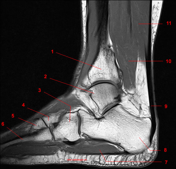
Plantar Tendons Of The Foot Mr Imaging And Us Radiographics from pubs.rsna.org Almost every muscle constitutes one part of a pair of identical bilateral. This is a table of skeletal muscles of the human anatomy. There are around 650 skeletal muscles within the typical human body. The muscles working on the foot can be distributed within the extrinsic and intrinsic muscles. Head, neck, arm, foot, pelvis, etc. There are 10 intrinsic muscles located in the sole of the foot. Common questions and answers about foot anatomy mri. Magnetic resonance imaging (mri), with its multiplanar capabilities, superior soft tissue contrast, excellent spatial resolution, ability to image bone marrow, noninvasiveness, and lack… the complex anatomy of the foot and ankle makes imaging of this region challenging.
The muscles that control the movements of the foot originate in the lower leg and are attached the bones in the foot with tendons.
Learn anatomy faster and remember everything you learn. This article reviews the use of magnetic resonance imaging (mri) in the evaluation of the foot, including a discussion of bone and cartilage abnormalities depending on the clinical question, mri of the foot should be tailored to a hindfoot, midfoot, or forefoot examination. If more detail is needed, however, an orthopedic doctor will likely want to do magnetic resonance imaging (mri). This article discusses the anatomy, supply, function and clinical relevance of the dorsal muscles of the foot. This is a table of skeletal muscles of the human anatomy. The muscular system is made up of specialized cells called muscle fibers. Radiologists perform ankle imaging to assess injuries of the foot and ankle anatomy. Attached to the bones of the skeletal system are about 700 named. The anatomy of the foot and common foot problems. Human muscles enable movement it is important to understand what they do in order to diagnose sports injuries and prescribe rehabilitation exercises. The feet are flexible structures of bones, joints, muscles, and soft tissues that let us stand upright and perform activities like walking, running, and jumping. The muscles lie within a flat fascia on the dorsum of the foot (fascia dorsalis pedis) and are innervated by the deep fibular or. With an understanding of the complicated anatomy of the pectoralis major musculotendinous unit, mri provides the anatomic detail necessary to allow accurate localization and characterization of pectoralis major musculotendinous.
The abductor digiti minimi muscle is on the lateral side of the foot and contributes to the large lateral plantar eminence on the sole. Their main function is contractibility. Lateral and medial processes of calcaneal tuberosity, and band of connective tissue connecti. Like the fingers, the toes have flexor and extensor muscles that power their movement and play a large role in balance. The muscles acting on the foot can be divided into two distinct groups;
Mri Of The Ankle Detailed Anatomy W Radiology from w-radiology.com Ankle and foot | radiology key. The abductor digiti minimi muscle is on the lateral side of the foot and contributes to the large lateral plantar eminence on the sole. Learn anatomy faster and remember everything you learn. 12 photos of the foot muscle anatomy mri. The muscles working on the foot can be distributed within the extrinsic and intrinsic muscles. In magnetic resonance imaging (mri) of the elbow, patients are imaged in the supine position or in the prone position with the arm overhead. Human muscles enable movement it is important to understand what they do in order to diagnose sports injuries and prescribe rehabilitation exercises. Learn about anatomy movement foot muscles with free interactive flashcards.
Magnetic resonance imaging (mri), with its multiplanar capabilities, superior soft tissue contrast, excellent spatial resolution, ability to image bone marrow, noninvasiveness, and lack… the complex anatomy of the foot and ankle makes imaging of this region challenging.
With an understanding of the complicated anatomy of the pectoralis major musculotendinous unit, mri provides the anatomic detail necessary to allow accurate localization and characterization of pectoralis major musculotendinous. Muscle anatomy diagram, dog muscle anatomy diagram, lower leg muscle anatomy diagram, muscle anatomy of human back, tricep muscle anatomy diagram, human muscles, canine muscle anatomy diagram, dog muscle anatomy. Learn anatomy faster and remember everything you learn. Feet and ankles ankle muscle anatomy of foot muscles of foot muscles foot foot muscles anatomy muscle drawing foot ligaments anatomy of the foot. Almost every movement in the body is the outcome of muscle contraction. Head, neck, arm, foot, pelvis, etc. Radiologists perform ankle imaging to assess injuries of the foot and ankle anatomy. Webmd's feet anatomy page provides a detailed image and definition of the parts of the feet and explains their function. Attached to the bones of the skeletal system are about 700 named. Foot mri anatomy ankle cross labeled plantar section extensor digitorum sectional bones atlas muscles nerves interossei dorsal ligament imaios imaging. Their main function is contractibility. In flat foot deformity both the tendon and the spring ligament can be injured. Pectoralis muscle mri & anatomy.
Tendinous, ligamentous, and muscle abnormalities. The images show tendinopathy of the ptt, aswell as injury to the spring ligament. There are 10 intrinsic muscles located in the sole of the foot. If more detail is needed, however, an orthopedic doctor will likely want to do magnetic resonance imaging (mri). Almost every movement in the body is the outcome of muscle contraction.
Anatomy Of The Foot And Ankle Mri from www.imaios.com Head, neck, arm, foot, pelvis, etc. Muscles, connected to bones or internal organs and blood vessels, are in charge for movement. Attached to the bones of the skeletal system are about 700 named. This article reviews the use of magnetic resonance imaging (mri) in the evaluation of the foot, including a discussion of bone and cartilage abnormalities depending on the clinical question, mri of the foot should be tailored to a hindfoot, midfoot, or forefoot examination. Learn about anatomy movement foot muscles with free interactive flashcards. The anatomy of the foot and common foot problems. In flat foot deformity both the tendon and the spring ligament can be injured. Ankle and foot | radiology key.
Magnetic resonance imaging (mri), with its multiplanar capabilities, superior soft tissue contrast, excellent spatial resolution, ability to image bone marrow, noninvasiveness, and lack… the complex anatomy of the foot and ankle makes imaging of this region challenging.
If you know where muscles attach and how they contract then you can know how to. Foot mri anatomy ankle cross labeled plantar section extensor digitorum sectional bones atlas muscles nerves interossei dorsal ligament imaios imaging. Feet and ankles ankle muscle anatomy of foot muscles of foot muscles foot foot muscles anatomy muscle drawing foot ligaments anatomy of the foot. Ankle and foot | radiology key. There are around 650 skeletal muscles within the typical human body. The foot is a part of vertebrate anatomy which serves the purpose of supporting the animal's weight and allowing for locomotion on land. Attached to the bones of the skeletal system are about 700 named. 12 photos of the foot muscle anatomy mri. Almost every movement in the body is the outcome of muscle contraction. Mri of the ankle and feet. Learn anatomy faster and remember everything you learn. The muscles that control the movements of the foot originate in the lower leg and are attached the bones in the foot with tendons. Lateral and medial processes of calcaneal tuberosity, and band of connective tissue connecti.
Feet and ankles ankle muscle anatomy of foot muscles of foot muscles foot foot muscles anatomy muscle drawing foot ligaments anatomy of the foot foot muscles mri. Like the fingers, the toes have flexor and extensor muscles that power their movement and play a large role in balance.


0 Komentar